- Abstract
- Introduction
- Materials and Methods
- Chemicals and reagents
- Instruments used
- Collection of insect samples and gut dissection
- Isolation and screening of lignin-degrading gut bacteria
- Genomic DNA extraction
- PCR amplification of the 16S rRNA gene
- Sequence similarity analysis
- Multiple sequence alignment
- Phylogenetic tree construction
- Criteria for species identification
- Results and Discussion
- Conclusion
- Funding
- Availability Statement
- AI Use Statement
- Authors Contributions
- Conflict of Interests
- References

Int J Chem Res, Vol 10, Issue 4, 19-24Research Article
Molecular Identification and Phylogenetic Characterization of Lignocellulose-Degrading Gut Bacteria Isolated From the Eri Silkworm (Samia cynthia ricini)
SHESHU M.*, MALLIAH SHIVASHANKAR
Department of Life Science, Bangalore University, Jnanabharathi, Jnana Jyothi Nagar, Gnana Bharathi, Karnataka-560056, Bengaluru, India
*Corresponding author: Sheshu M.; *Email: [email protected]
Received: 02 Jun 2026 Revised and Accepted: 22 Jul 2026
Abstract
Objective: The present study was aimed at identifying and characterising lignocellulose-degrading bacterial isolates from the gut of the eri silkworm, Samia cynthia ricini, by 16S rRNA gene sequencing and phylogenetic analysis.
Methods: Genomic DNA was isolated from six bacterial isolates and PCR amplified for the 16S rRNA gene. The amplified products were sequenced and aligned with reference sequences available in the NCBI GenBank database using BLAST analysis. Multiple sequence alignment was performed, and phylogenetic relationships were deduced using the Neighbour-Joining method with 1000 bootstrap replicates in MEGA11.
Results: Sequence analysis revealed that the isolates are Bacillus paralicheniformis (BUBSL1, 99.2%), Bacillus subtilis (BUBSL2, 99.5%), Bacillus licheniformis (BUBSL3, 98.9%), Paenibacillus sp. (BUBSL4, 97.8%), Bacillus velezensis (BUBSL5, 99.3%) and Paenibacillus lactis (BUBSL6, 99.0%). Phylogenetic analysis grouped the isolates into the genera Bacillus and Paenibacillus, supporting their taxonomic position and evolutionary relationship, and the clustering pattern showed that the isolates were closely related to the reference strains obtained from GenBank.
Conclusion: The gut microbiota of Samia cynthia ricini is composed of diverse bacterial species predominantly from the genera Bacillus and Paenibacillus. The isolated strains are potential lignocellulose-degrading bacteria, which can be used for future biofuel-related studies and may assist in biomass conversion.
Keywords: 16S rRNA gene, Phylogenetic analysis, Gut microbiota, Lignocellulose degradation, Eri silkworm, Bacillus, Paenibacillus
© 2026 The Authors.Published by Innovare Academic Sciences Pvt Ltd. This is an open access article under the CC BY license (http://creativecommons.org/licenses/by/4.0/)
DOI: http://dx.doi.org/10.22159/ijcr.2026v10i4.360 Journal homepage: https://ijcr.info/index.php/journal
Introduction
Lignocellulosic biomass is considered to have one of the most abundant and renewable organic resources available for sustainable energy production. The material is mainly composed of cellulose, hemicellulose, and lignin, which form complex hybrid structures that are very difficult to degrade. Lignin is one of the major components that contribute to the structure, forming a barrier that provides rigidity and protects plant cell walls; its presence limits enzyme access to polysaccharides and reduces the efficiency of biomass conversion [1, 2]. Microorganisms capable of degrading lignocellulosic materials are of great interest because they are known to produce a wide range of hydrolytic and oxidative enzymes, which are crucial for the breakdown of complex plant polymers into simple compounds that can be used for biofuel production and other industrial applications [3]. Bacterial systems have been promising in this aspect due to their rapid growth and ability to adapt in diverse environmental conditions and produce enzymes in large quantities. Insect gut ecosystems have recently been recognised as important reservoirs for lignocellulose-degrading microorganisms. Recent metagenomic and culture-dependent studies have further demonstrated that insect gut microbiota constitute valuable reservoirs of lignocellulolytic microorganisms with significant potential for biomass valuation and industrial biotechnology applications. Herbivorous insects depend on symbiotic microbial communities to digest a complex diet of plant polymers. Microbes are essential in breaking down lignocellulosic substrates, thereby increasing the bioavailability of nutrients to the host and its metabolism [4, 5]. The insect gut environment can thus be regarded as a natural bioconversion system for efficient degradation of lignocellulose under mild physiological conditions, such as low pH and temperature. For example, lepidopteran larvae are known to harbor complex microbial communities, which is important for the degradation of plant materials. The eri silkworm, Samia cynthia ricini, feeds on leaves rich in lignocellulose, and hence its gut is a potential niche for the isolation of bacteria with lignin-degrading potential. Interactions between the gut microbiota and the host organism are required for efficient digestion, nutrient uptake and survival. Bacterial genera such as Bacillus and Paenibacillus have been reported in the insect gut environment, and these genera are known for their production of cellulases, hemicellulases, and lignin-modifying enzymes, which are important for the degradation of plant biomass [6, 7]. However, molecular characterisation and phylogenetic analysis of lignocellulose-degrading bacteria from the guts of Samia cynthia ricini are still lacking. The correct bacterial species identification is important for understanding their ecological roles and biotechnological applications. The conserved and variable regions of the 16S rRNA gene are crucial for bacterial identification. These methods are extensively used as molecular approaches for identifying bacterial species and can discriminate between bacteria at different taxonomic levels [8]. Moreover, phylogenetic analysis provides the evolutionary relationship and facilitates the identification at the species level [9, 10]. The objective of the study was to isolate and identify lignin-degrading bacteria from the gut of Samia cynthia ricini by using 16S rRNA gene sequencing. Recent progress in microbial ecology and biofuel biotechnology has suggested that Bacillus and Paenibacillus spp. represent promising candidates for the degradation of lignocellulose and sustainable bioenergy production [24]. Phylogenetic analysis was performed to establish evolutionary relationships and confirm taxonomic placement. This study aims to help clarify the insect gut microbiota and its potential application in lignocellulose degradation and biofuel production.
Materials and Methods
Chemicals and reagents
All chemicals and reagents used in the present study were of analytical grade. Nutrient agar (NA), nutrient broth (NB), agarose, ethidium bromide, Tris-EDTA (TE) buffer, phosphate-buffered saline (PBS), sodium chloride (NaCl), molecular biology-grade water and DNA loading dye were purchased from HiMedia Laboratories Pvt. Ltd, Mumbai, India. PCR master mix, Taq DNA polymerase, dNTPs, universal 16S rRNA primers (27F and 1492R) and DNA molecular weight markers (1 kb DNA ladder) were procured from Thermo Fisher Scientific, USA. DNA extraction reagents and purification kits were purchased from Qiagen, Germany. Ethanol (70%) and other routine laboratory reagents were obtained from Merck Life Science Pvt. Ltd., India.
Instruments used
All experimental procedures were carried out using standard microbiological and molecular biology equipment. Sterile manipulations were performed in a laminar airflow cabinet (Ozone Scientific, Bengaluru, Karnataka, India). The bacterial cultures were incubated in a BOD incubator (Sunrise Enterprises, Bengaluru, Karnataka, India). The cell harvesting and processing of the samples was done with the help of a refrigerated centrifuge (REMI R-4C DX table Top Centrifuge; Remi Elektrotechnik Ltd., Mumbai, Maharashtra, India). The 16S rRNA gene was amplified by a thermal cycler (Veriti™ 96, Thermo Fisher Scientific, Waltham, MA, USA). The amplified DNA fragments were separated by agarose gel electrophoresis using a horizontal electrophoresis unit and visualised by a gel documentation system (Thermo Fisher Scientific, Waltham, Massachusetts, USA). DNA concentration and purity were determined using a UV–Visible spectrophotometer (Model 117, Systronics India Ltd, Ahmedabad, Gujarat, India). Sequencing of amplified 16S rRNA gene products was carried out by a commercial sequencing facility (Sakhala Enterprises, Rajarajeshwari Nagar, Bengaluru, Karnataka, India). All instruments were operated according to the manufacturer’s instructions.
Collection of insect samples and gut dissection
Healthy larvae of Samia cynthia ricini were obtained from a controlled rearing facility, Central Sericultural Germplasm Resources Centre (CSGRC), and transported to the laboratory under aseptic conditions. To remove any external microbial contaminants, the larvae were surface sterilised with 70% ethanol and subsequently rinsed multiple times with sterile distilled water. Dissection was done in a laminar airflow chamber under sterile conditions. The entire gut was surgically removed and placed in sterile phosphate-buffered saline (PBS). Gut tissues were then thoroughly homogenised to obtain a uniform microbial suspension for further isolation procedures [11].
Isolation and screening of lignin-degrading gut bacteria
The gut homogenate was serially diluted in sterile saline solution and plated on nutrient agar plates. The plates were incubated at 37 °C for 24–48 h. Colonies with different morphological characters were picked and purified by repeated streaking to obtain pure cultures. The purified isolates were inoculated on minimal salt medium with lignin (MSM-L) as the sole carbon source for selective screening. The composition of MSM-L (per litre) was as follows: KH₂PO₄, 1.0 g; K₂HPO₄, 1.0 g; NH₄NO₃, 1.0 g; MgSO₄·7H₂O, 0.2 g; CaCl₂, 0.02 g; FeSO₄·7H₂O, 0.01 g; NaCl, 0.5 g; and alkali lignin, 0.5–1.0% (w/v). The pH was adjusted to 7.0, and agar (15 g/l) was added for the solid medium. The inoculated plates were incubated at 30–37 °C for a period of 3–5 days. Growth on MSM-L was considered indicative of the ability of isolates to utilise lignin as a carbon source. Ligninolytic activity was also quantified by dye decolourisation assays, where the presence of clear or decolourised zones around colonies indicated enzymatic degradation of lignin-related compounds [12]. The isolates with positive activity were then analysed in a molecular approach [1, 7].
Genomic DNA extraction
Pure bacterial isolates were grown in nutrient broth overnight for harvesting enough biomass. Cells were collected by centrifugation, and genomic DNA was extracted using a modified phenol–chloroform method. Cell pellets were lysed using an appropriate lysis buffer, and proteins were digested by proteinase K. DNA was purified by phenol-chloroform extraction and precipitated by cold ethanol. The quality and integrity of the extracted DNA were checked by agarose gel electrophoresis, and the concentration was determined spectrophotometrically [13].
PCR amplification of the 16S rRNA gene
The 16S rRNA gene was amplified using universal bacterial primers, which bind to the conserved regions of the gene. PCR amplification was carried out on a thermal cycler with optimised reaction conditions, which consisted of an initial denaturation step followed by repeated cycles of denaturation, primer annealing and extension and the final extension step. The amplified products were separated on 1% agarose gel and visualised under UV light. The presence of a single band of ~ 1400 bp indicated the successful amplification. The 16S rRNA gene has been widely used for bacterial identification, as it contains conserved as well as variable regions, which provide taxonomic resolution. Sequencing and submission to GenBank. The purified PCR products were sequenced by the Sanger method, and the sequences were edited and assembled by bioinformatics tools to remove ambiguities and confirm their accuracy. The high-quality sequences were submitted to the NCBI GenBank database, and accession numbers were assigned for each isolate. The high-quality 16S rRNA gene sequences were deposited in the NCBI GenBank database and assigned publicly accessible accession numbers (PX884302, PX904987, PX904988, PX904993, PX904997 and PX904999), ensuring transparency, reproducibility, and future accessibility of the sequence data [14].
Sequence similarity analysis
The 16S rRNA gene sequences obtained were compared with the reference sequences available in the NCBI database by the Basic Local Alignment Search Tool (BLAST). Percentage similarity values were calculated in order to find the closely related species. BLAST analysis is a common method to determine rapid and reliable identification of microbial sequences based on nucleotide homology [15].
Multiple sequence alignment
Sequences with high similarity were selected and aligned to the reference sequences using the Clustal-W algorithm. Multiple sequence alignment was performed using approximately 799 bp of partial 16S rRNA gene sequences. Multiple sequence alignment allowed identification of conserved and variable nucleotide regions that are important for accurate phylogenetic analysis [16].
Phylogenetic tree construction
Phylogenetic relationships among the isolates and reference strains were analysed by the neighbour-joining method implemented in MEGA11 software [17]. Evolutionary distances among sequences were calculated using the Kimura two-parameter model, which takes into account both transition and transversion substitution rates [18]. The branching points were tested by bootstrap analysis with 1000 replicates [19, 20]. An appropriate outgroup was used to root the tree and interpret the evolutionary relationship. Closely related reference sequences and valid type strains retrieved from the GenBank database were included in the phylogenetic reconstruction to confirm species-level assignment and evolutionary relationships.
Criteria for species identification
Species-level identification was assigned when 16S rRNA gene sequence similarity was ≥98.7% and the isolate clustered with the corresponding reference species in the phylogenetic tree. Isolates showing similarity below 98.7% were identified only to the genus level [9, 10].
Results and Discussion
Molecular identification of isolates
All six bacterial isolates (BUBSL1-BUBSL6) showed clear and discrete bands of approximately 1400 base pairs, indicating successful amplification of the 16S rRNA gene. The sharpness and uniformity of the bands indicated the quality of genomic DNA as well as the efficiency of the PCR parameters employed.
Amplified products were sequenced, and the nucleotide sequences obtained were used in similarity searches with the BLAST algorithm. The analysis showed that all the isolates had a high similarity (97-99%) with previously reported bacterial species. Most of the isolates showed closest affiliation with members of the genus Bacillus, while two isolates showed strong similarity to Paenibacillus species. The high similarity values (>97%) suggest a reliable taxonomic affiliation between the isolates and previously characterised species. The sequences from this study were submitted to the GenBank database to ensure accessibility and reproducibility of the molecular data. The predominance of Bacillus and Paenibacillus suggests that these genera are well adapted to the gut environment of Samia cynthia ricini, especially for the degradation of lignocellulosic substrates.
Table 1: BLAST-based identification and GenBank accession numbers of bacterial isolates.
| Isolate | Closest match | Similarity (%) | Accession number |
| BUBSL1 | Bacillus paralicheniformis | 99.2 | PX884302 |
| BUBSL2 | Bacillus subtilis | 99.5 | PX904987 |
| BUBSL3 | Bacillus licheniformis | 98.9 | PX904988 |
| BUBSL4 | Paenibacillus sp. | 97.8 | PX904993 |
| BUBSL5 | Bacillus velezensis | 99.3 | PX904997 |
| BUBSL6 | Paenibacillus lactis | 99.0 | PX904999 |
Identification of bacterial isolates was based on BLASTn analysis of partial 16S rRNA gene sequences against the NCBI GenBank database. Sequence similarity values represent the closest matching reference sequences. GenBank accession numbers correspond to publicly accessible records deposited by the authors. Isolate BUBSL4 exhibited 97.8% sequence similarity and was therefore assigned only to the genus level as Paenibacillus sp.
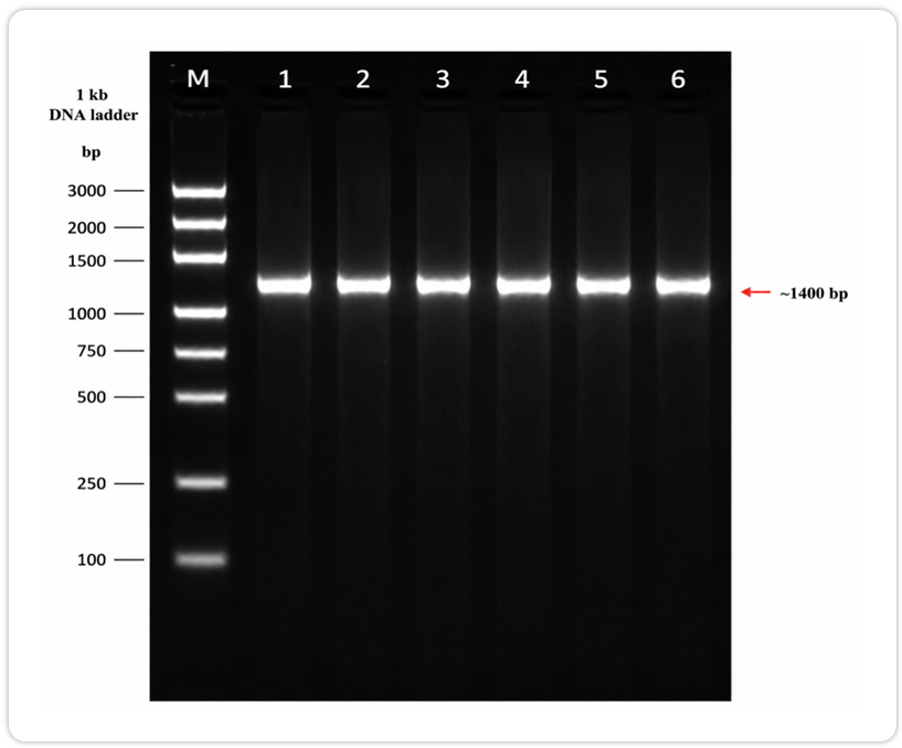
Fig. 1: Agarose gel electrophoresis of amplified 16S rRNA gene fragments. M = 1 kb DNA ladder. Lanes 1–6 correspond to isolates BUBSL1–BUBSL6. A distinct amplicon of approximately 1400 bp was observed in all isolates. M represents a 1 kb DNA ladder used as a molecular size marker. Lanes 1–6 correspond to isolates BUBSL1–BUBSL6, respectively. A single amplification product of approximately 1400 bp was obtained for all isolates using universal bacterial 16S rRNA gene primers.
Phylogenetic analysis
For further confirmation of the taxonomic identity of the isolates, phylogenetic analysis was performed using the Neighbour-Joining method. Evolutionary distances were computed with the Kimura two-parameter model that considers the differences in the rates of nucleotide substitutions and provides a reliable estimate of the genetic divergence [18]. The most widely used method to infer phylogenetic relationships is the Neighbour-Joining method because of its speed and accuracy. The phylogenetic tree obtained clustered the isolates into two groups, corresponding to the genera Bacillus and Paenibacillus [19]. The clustering pattern was in accordance with the results of the similarity BLAST, thus confirming the reliability of the molecular identification. The tree was subjected to bootstrap analysis with 1000 replicates. Several major branches were supported moderately to highly (>70%), whereas some internal nodes had lower support values, indicating uncertainty in some of the branching relationships. High bootstrap values indicate that the grouping of the isolates is stable and reflects true evolutionary relationships and not random association. The addition of suitable reference sequences and an outgroup also made the phylogenetic tree properly rooted. The separation of Bacillus and Paenibacillus into separate clades is in agreement with the bacterial taxonomy and divergence [21].
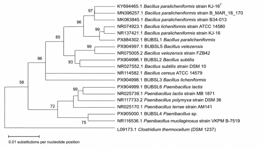
Fig. 2: Neighbour-joining phylogenetic tree based on partial 16S rRNA gene sequences showing the evolutionary relationships among six bacterial isolates and fourteen closely related reference strains retrieved from GenBank. A total of 20 taxa were analysed using approximately 799 aligned nucleotide positions. Evolutionary distances were calculated using the Kimura two-parameter model in MEGA11. Branch support values were estimated using 1000 bootstrap replicates and are shown at the corresponding nodes. Clostridium thermocellum DSM 1237 was used as the outgroup to root the tree. The scale bar represents 0.01 substitutions per nucleotide position. Closely related reference sequences and valid type strains retrieved from GenBank were included in the phylogenetic reconstruction. Bootstrap support values obtained from 1000 replicates are shown at major nodes value under 50% were omitted for clarity.
Species-level identification
The assignment of BUBSL6 to Paenibacillus lactis was supported by both BLAST similarity (99.0%) and phylogenetic clustering with reference P. lactis strains. The isolates were identified to the species level based on combined evidence from sequence similarity. Phylogenetic analysis revealed that the six bacterial isolates belonged to two major genera, Bacillus and Paenibacillus. BUBSL1 was isolated and showed close affiliation with Bacillus paralicheniformis. BUBSL2 clustered with Bacillus subtilis. BUBSL3 clustered with Bacillus licheniformis and BUBSL5 showed a close relationship with Bacillus velezensis. Isolate BUBSL4 clustered within the Paenibacillus clade but could not be assigned confidently to a particular species because of its lower sequence similarity and was therefore designated as Paenibacillus sp. In contrast, BUBSL6 clustered closely with Paenibacillus lactis. The observed clustering pattern was consistent with the results of BLAST-based identification and further confirmed the taxonomic assignment of the isolates. As indicated in the phylogenetic tree, the clustering pattern strongly supports the taxonomical position of the isolates. Reliable bacterial identification with high accuracy can be achieved using phylogenetic analysis and sequence similarity threshold methods [9, 10].
Table 2: Phylogenetic classification of bacterial isolates based on 16S rRNA analysis.
| Isolate | Genus | Species |
| BUBSL1 | Bacillus | B. paralicheniformis |
| BUBSL2 | Bacillus | B. subtilis |
| BUBSL3 | Bacillus | B. licheniformis |
| BUBSL4 | Paenibacillus | Paenibacillus sp. |
| BUBSL5 | Bacillus | B. velezensis |
| BUBSL6 | Paenibacillus | P. lactis |
Species assignments were supported by both BLAST sequence similarity and phylogenetic clustering with closely related reference strains retrieved from GenBank. Closely related type strains were included in the phylogenetic reconstruction to improve taxonomic resolution.
Functional significance of the identified isolates
The functional relevance of detected isolates, Bacillus and Paenibacillus species, in the gut of Samia cynthia ricini is indicative of the role of these microorganisms in lignocellulose degradation. The Bacillus genus is known to produce a wide range of extracellular enzymes, such as cellulases, xylanases and lignin-modifying enzymes [6], participating in the breakdown of plant biomass.
Particularly noteworthy was the presence of B. subtilis and B. velezensis, two species that are characterised by high enzyme activity and adaptability to different environmental conditions. Bacillus licheniformis produces enzymes of industrial importance for the degradation of lignocellulose and bioconversion of biomass. The genus Paenibacillus is reported to have a wide range of carbohydrate-active enzyme systems for the degradation of complex polysaccharides. The presence of P. lactis and Paenibacillus sp. supports the hypothesis that different groups of bacteria act synergistically in the degradation of lignocellulose in the gut environment. Recent genomic and phylogenetic investigations have similarly reported the involvement of Paenibacillus species in plant biomass degradation and carbohydrate metabolism [24].
Comparison with other studies
Consistent with other studies on insect gut microbiota, the results of this study have shown that the dominant bacteria in lignocellulose degradation are Bacillus and Paenibacillus spp. Insect digestive systems contain microbial groups that work together to break down complex plant molecules, thus improving the nutritional quality for the host [4, 5]. Bacillus species are relevant in cellulose degradation and nutrient assimilation in other lepidopteran insects, and similar microbial profiles have been reported. The presence of these bacteria in the gut of Samia cynthia ricini corroborates the concept of insect guts as natural bioreactors for lignocellulose conversion [1, 23]. Moreover, the studies of the erisilkworm gut microbiota showed that bacterial symbionts have a crucial role in digestive and metabolic processes, demonstrating the ecological role of these microorganisms for the host’s survival and adaptation.
General discussion
Our molecular identification and phylogenetic analysis provide a strong and comprehensive approach for the study of the diversity of gut-associated bacteria in Samia cynthina ricini. The isolates were found to be in clusters of Bacillus and Paenibacillus with high bootstrap support, and this result confirms the position of the isolates in taxonomy and evolutionary relationships. The ability of the isolates to grow in a medium supplemented with lignin shows their functional relevance in lignocellulose degradation. These results indicate that the gut microbiota of Samia cynthia ricini has a specific ability to degrade complex substrates of plant origin efficiently. Bacillus and Paenibacillus species produce cellulases, xylanases, laccases, and other lignocellulolytic enzymes. These species in the gut of Samia cynthia ricini may therefore act synergistically to degrade the lignocellulosic substrates eaten by the host insect.
Implications and future perspectives
The present study provides important knowledge about the diversity and functional potential of lignocellulose-degrading bacteria associated with the gut of Samia cynthia ricini. Further studies on enzyme characterisation, metabolic profiling, and genome analysis will improve our understanding of the mechanisms of lignocellulose degradation in these bacteria and their use in industrial biotechnology.
Novelty of the study
To our knowledge, this study is the first report describing the molecular identification and phylogenetic characterisation of lignocellulose-degrading gut bacteria associated with Samia cynthia ricini from India. Identification of several lignocellulolytic Bacillus and Paenibacillus species presents novel insights into the microbial ecology of the eri silkworm gut and its possible use in biomass valorisation and biofuel generation.
Conclusion
Six lignocellulolytic bacterial isolates were isolated from the gut of Samia cynthia ricini through 16S rRNA gene sequencing and phylogenetic analysis in this study. The isolates were identified as Bacillus paralicheniformis, Bacillus subtilis, Bacillus licheniformis, Bacillus velezensis, Paenibacillus sp., and Paenibacillus lactis. Their existence in the silkworm gut shows the ecological importance of gut microbiota in the degradation of lignocellulose. The bacterial isolates are promising candidates for future studies on enzyme production, biomass conversion and bioethanol production, supporting recent efforts to develop sustainable microbial platforms for renewable biofuel generation. These findings demonstrate the potential of the eri silkworm gut microbes as a source of lignocellulolytic bacteria for future biomass conversion and bioethanol production.
Funding
Nil
Availability Statement
The datasets generated and/or analyzed during the current study are available from the corresponding author on reasonable request.
AI Use Statement
The authors used ChatGPT (OpenAI) and Quillbot only for language editing and improving the readability of the manuscript. There was no use of AI tools in the data generation, analysis, interpretation, or preparation of scientific conclusions. The authors have read and checked all contents and take full responsibility for the manuscript.
Authors Contributions
Sheshu M. conceptualised the extensive literature, critically revised the manuscript, corrected and interpreted the data, and created graphical content and figures.
Malliah Shivashankar provided guidance in structuring the critical evolution of content and reviewed and refined the final version of the manuscript for submission.
Conflict of Interests
Declared none
References
Lynd LR, Weimer PJ, Van Zyl WH, Pretorius IS. Microbial cellulose utilization: fundamentals and biotechnology. Microbiol Mol Biol Rev. 2002;66(3):506-77. doi: 10.1128/MMBR.66.3.506-577.2002, PMID 12209002.
Kumar R, Singh S, Singh OV. Bioconversion of lignocellulosic biomass: biochemical and molecular perspectives. J Ind Microbiol Biotechnol. 2008;35(5):377-91. doi: 10.1007/s10295-008-0327-8, PMID 18338189.
Bugg TD, Ahmad M, Hardiman EM, Rahmanpour R. Pathways for degradation of lignin in bacteria and fungi. Nat Prod Rep. 2011;28(12):1883-96. doi: 10.1039/C1NP00042J, PMID 21918777.
Engel P, Moran NA. The gut microbiota of insects – diversity in structure and function. FEMS Microbiol Rev. 2013;37(5):699-735. doi: 10.1111/1574-6976.12025, PMID 23692388.
Brune A. Symbiotic digestion of lignocellulose in termite guts. Nat Rev Microbiol. 2014;12(3):168-80. doi: 10.1038/nrmicro3182, PMID 24487819.
Schallmey M, Singh A, Ward OP. Developments in the use of Bacillus species for industrial production. Can J Microbiol. 2004;50(1):1-17. doi: 10.1139/W03-076, PMID 15052317.
Catalao MJ, Gil F, Moniz-Pereira J, Sao-Jose C, Pimentel M. Diversity in bacterial lysis systems: bacteriophages show the way. FEMS Microbiol Rev. 2013;37(4):554-71. doi: 10.1111/1574-6976.12006, PMID 23043507.
Lane DJ. 16S/23S rRNA sequencing. In: Stackebrandt E, Goodfellow M, editors. Nucleic acid techniques in bacterial systematics. New York: John Wiley & Sons; 1991. p. 115-75.
Stackebrandt E, Goebel BM. Taxonomic note: a place for DNA-DNA reassociation and 16S rRNA sequence analysis in the present species definition in bacteriology. Int J Syst Evol Microbiol. 1994;44(4):846-9. doi: 10.1099/00207713-44-4-846.
Chun J, Oren A, Ventosa A, Christensen H, Arahal DR, Da Costa MS. Proposed minimal standards for the use of genome data for the taxonomy of prokaryotes. Int J Syst Evol Microbiol. 2018;68(1):461-6. doi: 10.1099/ijsem.0.002516, PMID 29292687.
Cappuccino JG, Sherman N. Microbiology: a laboratory manual. 10th ed. Boston: Pearson; 2014.
Pointing SB. Qualitative methods for the determination of lignocellulolytic enzyme production by fungi. Fungal Divers. 1999;2:17-33.
Green MR, Sambrook J. Molecular cloning: a laboratory manual. 4th ed. Cold Spring Harbor: Cold Spring Harbor Laboratory Press; 2012.
Altschul SF, Madden TL, Schaffer AA, Zhang J, Zhang Z, Miller W. Gapped BLAST and PSI-BLAST: a new generation of protein database search programs. Nucleic Acids Res. 1997;25(17):3389-402. doi: 10.1093/nar/25.17.3389, PMID 9254694.
Thompson JD, Higgins DG, Gibson TJ. Clustal W: improving the sensitivity of progressive multiple sequence alignment through sequence weighting, position-specific gap penalties and weight matrix choice. Nucleic Acids Res. 1994;22(22):4673-80. doi: 10.1093/nar/22.22.4673, PMID 7984417.
Tamura K, Nei M, Kumar S. Prospects for inferring very large phylogenies by using the neighbor-joining method. Proc Natl Acad Sci USA. 2004;101(30):11030-5. doi: 10.1073/pnas.0404206101, PMID 15258291.
Tamura K, Stecher G, Kumar S. MEGA11: molecular evolutionary genetics analysis version 11. Mol Biol Evol. 2021;38(7):3022-7. doi: 10.1093/molbev/msab120, PMID 33892491.
Saitou N, Nei M. The neighbour-joining method: a new method for reconstructing phylogenetic trees. Mol Biol Evol. 1987;4(4):406-25. doi: 10.1093/oxfordjournals.molbev.a040454.
Espadinha D, Sobral RG, Mendes CI, Meric G, Sheppard SK, Carrico JA. Distinct phenotypic and genomic signatures underlie contrasting pathogenic potential of Staphylococcus epidermidis clonal lineages. Front Microbiol. 2019;10:1971. doi: 10.3389/fmicb.2019.01971, PMID 31507574.
Kumar V, Chhabra D, Shukla P. Advances in microbial cellulases and their industrial applications. Enzyme Res. 2012;2012:280696. doi: 10.1155/2012/280696.
Singh R, Kumar M, Mittal A, Mehta PK. Microbial enzymes: industrial progress in 21st century. 3 Biotech. 2016;6(2):174. doi: 10.1007/s13205-016-0485-8, PMID 28330246.
Masmoudi F, Alsafran M, Jabri HA, Hosseini H, Trigui M, Sayadi S. Halobacteria-based biofertilizers: a promising alternative for enhancing soil fertility and crop productivity under biotic and abiotic stresses-a review. Microorganisms. 2023;11(5):1248. doi: 10.3390/microorganisms11051248, PMID 37317222.
Vojnovic S, Aleksic I, Ilic-Tomic T, Stevanovic M, Nikodinovic-Runic J. Bacillus and Streptomyces spp. as hosts for production of industrially relevant enzymes. Appl Microbiol Biotechnol. 2024;108(1):185. doi: 10.1007/s00253-023-12900-x, PMID 38289383.
Grady EN, Mac Donald J, Liu L, Richman A, Yuan ZC. Current knowledge and perspectives of Paenibacillus: a review. Microb Cell Fact. 2016;15(1):203. doi: 10.1186/s12934-016-0603-7, PMID 27905924.